
Margaux Loyer
Diploma in Beauty and Massage Therapy, Certificates in Deep Tissue and Myofascial Release, RYT 200 Yoga Teacher, Bachelor of Health Science in Acupuncture
The skin’s role goes beyond just being a shield from external elements. The skin is deeply involved in our emotional, cognitive, and immune responses.
With its vast network of mechanoreceptors, sensory cells, and immune cells, the skin is in constant communication with the brain, acting as a bridge between the physical and emotional self.
The Brain-Skin Axis: A Two-Way Connection
The connection between the brain and skin is far deeper than we often recognise. Known as the brain-skin axis, this two-way communication pathway reflects the intricate relationship between our emotional states and skin health.
Skin rash or wounds stimulate the skin, causing pain and triggering a cascade of events that send signals to the brain, activating a stress response.
RELATED — Understanding Stress: The Silent Killer
Similarly, therapeutic touch like massage stimulates the skin in a positive way, sending signals to the brain that promote emotional and psychological well-being, releasing feel-good hormones such as serotonin, dopamine, and endorphins.[1]
This bidirectional flow between the brain and the skin illustrates the critical role the skin plays, not just as a physical barrier, but as an active communicator in both physical health and emotional regulation.
Brain imaging studies show that psychological stress alters the amygdala and hippocampus, potentially worsening stress-related skin conditions.
Under stress, the skin engages with immune cells, hormones, and neurotransmitters, regulating inflammation through pro- and anti-inflammatory pathways. This highlights the skin’s neuroendocrine role in the body’s stress response.[2]
Stress can disrupt the delicate equilibrium of immune cells and inflammatory mediators within the skin, leading to immune dysregulation and increased sensitivity to various skin disorders.
Psychological stress can worsen stress-related skin conditions
Additionally, stress adversely impacts skin barrier function, slow down wound healing, and stimulates the release of pro-inflammatory cytokines.
This can exacerbate existing conditions such as psoriasis, atopic dermatitis, acne and urticaria.
The Ectodermal Origin: The Skin and Brain Share Common Ground
A 2023 study examines the skin-brain axis, highlighting their shared ectodermal origin, which links skin and neurological health. Both skin and brain arise from the ectoderm in early development, creating a pathway for skin changes, like injury or inflammation, to impact brain function.[3]
Skin-to-skin contact, particularly in newborns, has long been recognised for its profound impact on early brain development and emotional health.
RELATED — Introduction to: Brain Health
Known as “kangaroo care,” this practice is especially important for premature infants who often miss crucial in-utero sensory experiences.
Recent research from Stanford Medicine demonstrates that increased skin-to-skin time significantly improves neurodevelopment. The study found that for every additional 20 minutes of skin-to-skin contact daily, preterm infants experienced a 10-point increase in neurodevelopmental scores at 12 months, showing the positive correlation between skin stimulation and cognitive development.[4,5]
Skin-to-skin contact not only provides crucial sensory input but also stimulates the release of hormones such as oxytocin, the so-called “bonding hormone,” which promotes emotional regulation and strengthens the parent-child bond.
The influence of skin stimulation on brain development extends beyond infancy. Throughout life, physical touch continues to play a critical role in emotional and cognitive functions, reinforcing the concept of the skin as a “third brain”.
From childhood into adulthood, tactile interactions can:
- enhance emotional stability
- improve mood
- regulate stress
These findings reveal that skin is more than a protective barrier; it actively supports mental health, emphasizing the value of touch therapies like massage.
Keratinocytes and the Immune Response: Skin as an Immunological Hub
While the skin serves as a sensory organ, it also plays a critical role in immune defense. It acts as the first line of defense against pathogens.
The key immune cells in the epidermis are epidermal dendritic cells (Langerhans cells) and Keratinocytes. These cells recognise and respond to environmental threats through various mechanisms, including the release of signaling molecules that influence inflammation and healing processes.[6]
When the skin is activated through pressure, UV light, temperature, or a sting for instance, these immune cells can trigger systemic responses, affecting not just local inflammation but also hormonal responses in the body.
Keratinocytes are the most common cells in the epidermis. They are the first line of defense against germs and infections. They can detect harmful substances by using special sensors called Toll-like receptors (TLRs). When they find something harmful, they trigger an immune response to help fight it off.
Keratinocytes also communicate with other immune cells in a few important ways. Such as:
- By producing antimicrobial peptides, which are small proteins that can kill germs
- By releasing signaling molecules (called cytokines, like interleukin–1β) that send messages to other immune cells to help respond to infections
- By creating chemokines, which are substances that attract other immune cells to the skin to help fight off invaders
- By directly activating certain immune cells, like T lymphocytes and natural killer (NK) cells, which are important for fighting infections
This communication helps the skin protect itself and respond to potential threats.[6]
Denda’s 2015 study shows that epidermal keratinocytes function like neurons, responding to stimuli by releasing molecules such as cortisol and oxytocin.[7]
Epidermal keratinocytes function like neurons
They detect mechanical stress, temperature, and chemical changes, positioning the skin as a “third brain” and its role in stress regulation, mood, immune function, and mental health.[7]
Mechanoreceptors and Sensory Pathways
In the skin we also have different sensory receptors, such as mechanoreceptors, that communicate with the brain in response to touch.
There are four main types of mechanoreceptors:
- Merkel cells
- Meissner’s corpuscles
- Ruffini endings
- Pacinian corpuscles
Each responds to different types of touch sensations, such as light touch, vibration, pressure, and temperature.
For example, when someone touches our skin, mechanoreceptors in our skin directly detect this mechanical stimulus and generate electrical signals (action potentials) that are transmitted to the spinal cord and then to the brain.
When the brain receives these signals, it responds by releasing its own neurotransmitters, such as serotonin, dopamine, and norepinephrine.[8]
This interaction shows how touch impacts the brain through skin stimulation.
In the study by Witzel et al. (2011), magnetoencephalography (MEG) was used to map brain responses during 15 minutes of electroacupuncture (EA) and acupressure (AP).
Both treatments stimulated the brain, with EA showing a stronger, more immediate effect. While acupressure’s impact was slower and more diffuse, it still activated the brain, demonstrating that skin stimulation, whether through mechanical or electrical means, communicates with the nervous system and influences brain activity.[9]
(a) For all subjects, the primary sources localized to the contralateral SI for EA (white dots) and AP (blue dots)
(b) Average time-courses for all trials of EA and AP (black, dashed lines) demonstrated analogous M20, M30, M50/M60 and M120 (those of AP being slightly delayed). Five minute subaverages (blue, red and green lines) show attenuation of peak-to-peak amplitude over the course of the run.
This demonstrates that simple tactile stimuli, such as acupressure, can influence neural responses associated with pain modulation and emotional regulation. Through the skin-brain axis, touch impacts our brain chemistry.
Discovery of Piezo Proteins: Translating Touch into Brain Activity
The Nobel Prize in Physiology in 2021 was awarded to David Julius and Ardem Patapoutian for their discovery of how the body senses temperature and touch.
Their work uncovered how specific proteins (TRPV1 and Piezo channels) play a role in detecting temperature and mechanical stimuli, respectively.
These proteins are essential for transmitting signals from the skin to the brain, and work in pairs with mechanoreceptors, informing us about our environment.
This system allows us to feel sensations such as warmth or pressure, essential for activities ranging from basic survival instincts to the pleasure derived from physical touch.
Without Piezo2, for example, we wouldn’t be able to feel something as simple as a light touch, which is crucial for sensing our environment and the pleasurable sensations we experience from interactions like a gentle massage. This discovery has broad implications, from understanding pain mechanisms to developing new treatments for chronic pain.[10]
Referencing
(1) Ascsillán, A. A., & Kemény, L. V. (2024). The Skin–Brain Axis: From UV and Pigmentation to Behaviour Modulation. International Journal of Molecular Sciences, 25(11), 6199. https://www.mdpi.com/1422-0067/25/11/6199
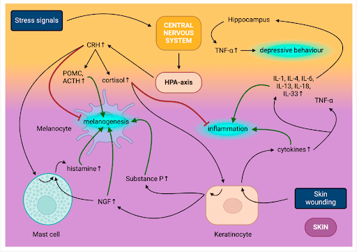
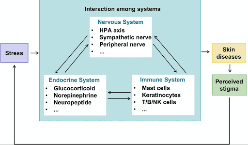
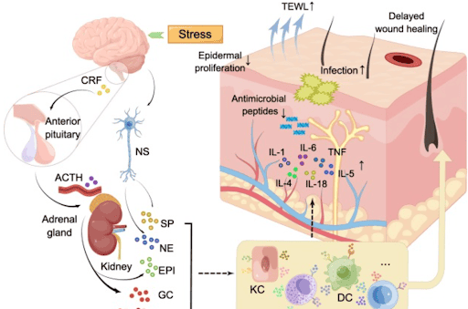
(2) Zhang, H., Wang, M., Zhao, X., Wang, Y., Chen, X., & Su, J. (2020). Role of stress in skin diseases: A neuroendocrine-immune interaction view. Journal of the European Academy of Dermatology and Venereology.
(3) Jameson, C., Boulton, K. A., Silove, N., Nanan, R., & Guastella, A. J. (2023). Ectodermal origins of the skin-brain axis: a novel model for the developing brain, inflammation, and neurodevelopmental conditions. Molecular Psychiatry, 28(1), 108–117. https://pmc.ncbi.nlm.nih.gov/articles/PMC9812765/
(4) Digitale, E. (2024, August 6). Skin-to-skin ‘kangaroo care’ found to boost neurodevelopment in preemies. Stanford Medicine.
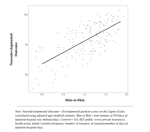
(5) Lazarus, M. F., Marchman, V. A., Brignoni-Pérez, E., Dubner, S., Feldman, H. M., & (2024). Inpatient Skin-to-Skin Care Predicts 12-month Neurodevelopmental Outcomes in Very Preterm Infants. medRxiv. https://www.medrxiv.org/content/10.1101/2023.04.06.23288260v2.full.pdf
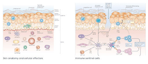
(6) Velykoredko, Y., Bohdanowicz, M., Oakley, A. (Editor-in-Chief), Mitchell, G. (Copy Editor), & McGivern, M. (Copy Editor). (2017). DermNet New Zealand. University of Toronto, Canada. https://dermnetnz.org/topics/skin-immune-system
(7) Denda, M. (2015). Epidermis as the “Third Brain”? Dermatologica Sinica, 33(2), 70–73.
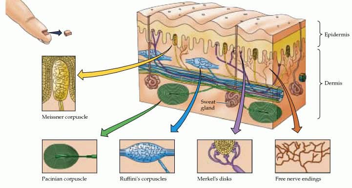
(8) Purves, D., Augustine, G. J., Fitzpatrick, D., et al. (2001). Mechanoreceptors specialized to receive tactile information. In Neuroscience (2nd ed.). Sunderland, MA: Sinauer Associates.
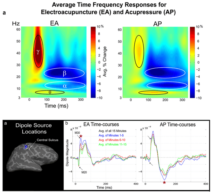
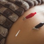
(9) Witzel, T., Napadow, V., Kettner, N. W., et al. (2011). Differences in cortical response to acupressure and electroacupuncture stimuli. BMC Neuroscience, 12, 73. https://bmcneurosci.biomedcentral.com/articles/10.1186/1471-2202-12-73
(10) Julius, D., & Patapoutian, A. (2021). Nobel Prize in Physiology or Medicine for the discovery of receptors for temperature and touch. NobelPrize.org. https://www.nobelprize.org/prizes/medicine/2021/press-release/